|
11/29/2021 0 Comments The Importance of a Pelvic Ultrasound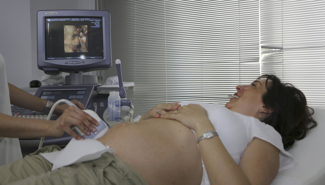 A pelvic ultrasound London can provide important information for women during pregnancy. This procedure can determine if a woman is carrying a fetus or not, and can also diagnose a range of problems relating to the uterus, ovaries, and endometrium. This procedure is not painful and takes about 30 minutes to complete. A woman will be asked to remove her clothes and lie down. If she spotting, she will need to remove the tampon before the scan. It can also be helpful to have a partially filled bladder, which helps to better see the organs in the pelvis. A private clinic in London will ask you to wear a gown and remove any clothing, which will also make the experience more comfortable. The ultrasound procedure itself does not require any anaesthetic. Patients will be asked to remove their underwear and lie on their back. This type of exam is accompanied by ultrasound gel which is applied to the skin to create high-resolution images of the pelvic area. Most patients will be able to hear the doctor's explanation of the results during the scan. The procedure is painless and does not leave any traces of ultrasound gel on their clothes. A specialist may refer you for a pelvic ultrasound in London if you're having pelvic pain. It usually takes less than 20 minutes and is far less invasive than a gynecological examination. It can also be a convenient way to get same-day appointments with specialists. A physician in London will be able to interpret the results of the procedure in a timely manner and recommend treatment. A woman may want to have a Private pelvic scan in London for a number of reasons. First of all, pelvic pain is usually a symptom of a gastrointestinal condition or a bowel disorder. The scan may reveal anything from urinary tract infection to breast cancer. If the symptoms are not present during the test, the doctor will be able to tell you. This test is often a valuable diagnostic tool. A woman only ultrasound is commonly performed on a woman and is performed internally. It can also be used to detect endometrial cancer, irregular bleeding, and inflammatory diseases of the pelvic region. However, it is not possible to guarantee the accuracy of the images. A doctor who performs a female pelvic ultrasound will need to use a transvaginal probe to do the scan. It is recommended that you emptied your bladder before the scan. The procedure is not painful. In fact, it can even be beneficial for women with a history of prostate cancer. Depending on the results, this examination may be beneficial for infertility. A patient will need to be fully conscious for the scan. The ultrasound will take pictures of the ovaries and uterus, and the doctor can use this information to help treat the patient. Further, this examination can also help diagnose some other conditions. Check out this related post to get more enlightened on the topic: https://en.wikipedia.org/wiki/Ultrasound.
0 Comments
11/29/2021 0 Comments Private Breast Ultrasound in London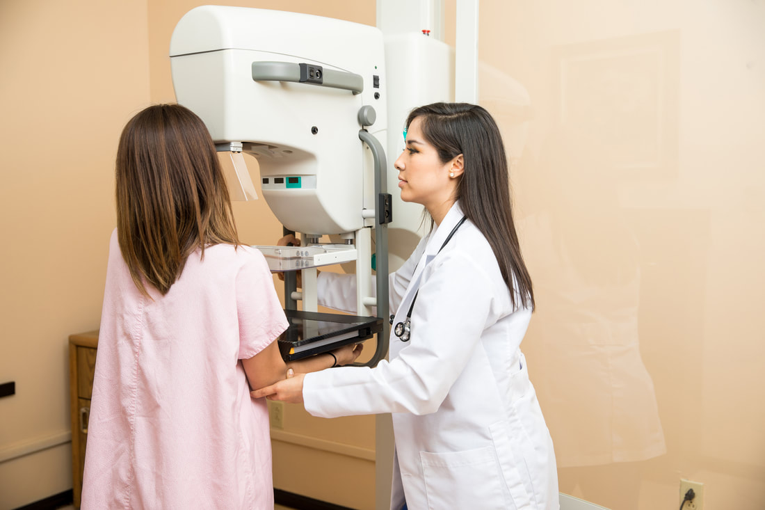 The best place for a private breast ultrasound in London is the Harley Street district. This medical centre is the choice of NHS consultants, local GPs and other health professionals. The clinic provides a comfortable, relaxed and professional environment for all diagnostic procedures, including breast ultrasound. The clinic also offers the most extensive range of private ultrasound examinations in the Harley Street area. The clinic has the highest patient satisfaction and patient referral rates in the UK. Breast ultrasound is a non-invasive procedure, involving lying on a bed and lifting one arm to the side. The technician will use a small ultrasound probe to take images of breast tissue. The breasts are then removed and the patient is asked to lie on a comfortable examination couch. No preparation is necessary and the treatment is completely painless. The patient's top and bra are removed, and there is no need to undress. A private breast ultrasound in London is a great way to check if your lump is cancerous or not. It is an essential step in breast cancer prevention and can help your doctor determine if you need further medical treatment. The results of the test will help your doctor determine whether you need to undergo a mammogram or breast ultrasound. If you notice a suspicious lump in your breast, an ultrasound is a good way to determine the source of the problem. Another way to find out if you have a mammogram is to go for an ultrasound. Mammograms are extremely common and are used to determine if a woman has breast cancer. However, an ultrasound can be much more accurate than a mammogram in many cases. A mammogram is a great diagnostic tool, but ultrasound is more detailed and may be a better option for younger women. A mammogram takes between 15 minutes and an hour, depending on the size of the breast. A Private ultrasound can be used to confirm a mammogram result. It can also help find out if there is a solid lump in the breast. It can also determine whether you have a solid mass in the breast. An ultrasound is an excellent way to find out whether you have a lump or a cyst in your breast. It can be helpful in determining the nature of a tumour, and can reveal the cause of a symptom. It is important to get a breast ultrasound if you suspect a lump. The procedure is an excellent way to discover whether a breast lump is a cancerous tumor. An ultrasound can also differentiate between solid and fluid-filled lumps and can detect a lump that is both. It is also crucial for women who are over 40 to get regular screenings for breast cancer. It can also help in detecting a symptom. Check out this related post to get more enlightened on the topic: https://en.wikipedia.org/wiki/Mammography. 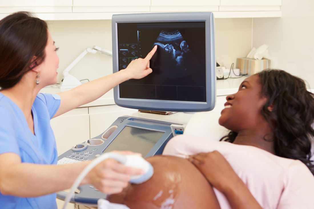 A pregnancy ultrasound is a medical test to determine whether the pregnancy is healthy or unhealthy. This test can be performed at any point in the pregnancy. The process is relatively safe, but the results can be affected by certain factors, including a woman's position. The positioning of the foetus can also affect the quality of the ultrasound. During your screening, the sonographer may ask you to move around a little, such as sitting in a particular position, or taking a different position for the next screening. A normal pregnancy ultrasound at 17 weeks gestation shows the placenta as a mound in the center of the screen. The uterus contracts periodically to provide better blood flow to the fetus. The nuchal translucency, a fluid sack at the back of the baby's neck filled with lymphatic fluid, may be visible at this time. If the uterus is not contracting, the placenta will be flattened out. A private pelvic ultrasound is a procedure that uses high-frequency sound waves to see the baby's organs. During this procedure, the woman is required to hold her breath for a few minutes and squat. A full bladder may cause some discomfort during the test, but this is usually very minimal. The doctor will then transform the echoes of the ultrasound into pictures on a computer screen. This type of scan takes about 30 minutes and the pictures are available for download. If the pregnancy ultrasound is performed at a hospital, it is recommended that you wear a maternity gown. The ultrasound technician will move the transducer across your belly and look at the fetus. The ultrasound technician will cover the transducer with a condom-like latex sheath to protect it from excessive pressure. In addition to this, you might have to take a few days off from work to change into a gown. In order to get the best images, the ultrasound technician moves the transducer over the pregnant woman's abdomen. In addition, he or she may take several minutes to get the perfect image. During the process, the ultrasound technician may be very quiet or speak only with the ultrasound technician. During the ultrasound, the baby's development is monitored through a series of images. The image shows the developing fetus' anatomy. A private scan during pregnancy is not recommended unless there is a medical reason for not using it. Despite the risks, pregnancy ultrasound is widely practiced in most obstetric units. However, pregnant women should not undergo the test unless they are aware of the risks. It is advisable for a woman to consult her doctor before getting pregnant. In general, it is a safe and healthy method. The procedure is not harmful for the mother or the baby. Check out this related post to get more enlightened on the topic: https://en.wikipedia.org/wiki/Ultrasound. |
AuthorWrite something about yourself. No need to be fancy, just an overview. ArchivesCategories |
 RSS Feed
